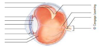
44 parts of the eye without labels
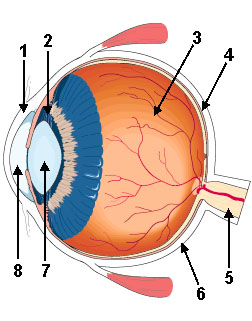
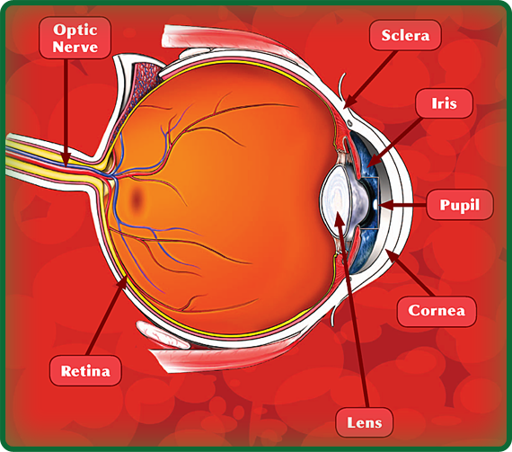
Labelling the eye — Science Learning Hub Labelling the eye. Use this interactive to label different parts of the human eye. Drag and drop the text labels onto the boxes next to the diagram. Selecting or hovering over a box will highlight each area in the diagram. The human eye has several structures that enable entering light energy to be converted to electrochemical energy. Anatomy of the Eye. Learn about the different parts of the eye. The sclera is a membrane of tendon in the eye, also known as the white of the eye. Rugged and robust, the sclera works to protect the inner, more sensitive parts of the eye like the retina and choroid. It is about 0.03 of an inch thick except for where the four "straight" eye muscles append, where the depth is no more than 0.01 of an inch.
Eye anatomy: A closer look at the parts of the eye Eye anatomy: A closer look at the parts of the eye. When surveyed about the five senses — sight, hearing, taste, smell and touch — people consistently report that their eyesight is the mode of perception they value (and fear losing) most. Despite this, many people don't have a good understanding of the anatomy of the eye, how vision works ...

Parts of the eye without labels
Anatomy of the Eye | Johns Hopkins Medicine Cornea. The clear, dome-shaped surface that covers the front of the eye. Iris. The colored part of the eye. The iris is partly responsible for regulating the amount of light permitted to enter the eye. Lens (also called crystalline lens). The transparent structure inside the eye that focuses light rays onto the retina. Lower eyelid. Eye in Cross Section : Anatomy : The Eyes Have It inner layer of posterior wall of eye (see Retina in Cross Section) contains receptors that convert light energy into signals that brain can interpret. Choroid: vascular layer that nourishes outer retina. can be inflamed in autoimmune ("rheumatologic") disorders. Sclera: collagenous outer layer of wall of eye. reference.yourdictionary.com › resources › parts-ofParts of the Body for Kids: Names & Basic Functions Diagram of Body Parts. External, which means “outside,” describes the body parts that you can see. Take a look at a helpful diagram that labels major external body parts. Download the printable PDF to see it in more detail and print if needed. View & Download PDF
Parts of the eye without labels. Label Parts of the Human Eye - University of Dayton Parts of the Eye Select the correct label for each part of the eye. The image is taken from above the left eye. Click on the Score button to see how you did. Incorrect answers will be marked in red. Quiz: Label The Parts Of The Eye - ProProfs How much did you get to understand about the human eye? Take up this quiz and find out! Questions and Answers. 1. A is pointing to what part of the eye? A. Cornea. B. Optic Nerve. Eye Pictures, Anatomy & Diagram | Body Maps - Healthline The eye has several major components: the cornea, pupil, lens, iris, retina, and sclera. These work together to capture an image and transmit it directly to the brain's occipital lobe via the optic... Eye Diagram Teaching Resources | Teachers Pay Teachers Use these simple eye diagrams to help students learn about the human eye. Three differentiated worksheets are included: 1. Write the words using a word bank2. Cut and paste the words3.
Anatomy of the Eye | Kellogg Eye Center | Michigan Medicine Structure containing muscle and is located behind the iris, which focuses the lens. Cornea The clear front window of the eye which transmits and focuses (i.e., sharpness or clarity) light into the eye. Corrective laser surgery reshapes the cornea, changing the focus. Fovea The center of the macula which provides the sharp vision. Iris Your Eyes (for Kids) - Nemours KidsHealth It is a very important part of the eye, but you can hardly see it because it's made of clear tissue. Like clear glass, the cornea gives your eye a clear window to view the world through. Iris Is The Colorful Part. Behind the cornea are the iris, the pupil, and the anterior chamber. The iris (say: EYE-riss) is the colorful part of the eye. When ... BYJUS BYJUS leafyplace.com › parts-of-a-flowerParts of Flower and Plant (Pistil, Sepal, Stamen and More ... Dec 26, 2019 · Extracts of many plants have medicinal properties that can help address a number of ailments. Parts of plants can be used to make therapeutic herbal teas, essential oils, or taken as supplements. Pleasing to the eye. Flowers, blossoms, and colorful petals are pleasing to the eye.
en.wikipedia.org › wiki › Human_eyeHuman eye - Wikipedia Each eye has seven extraocular muscles located in its orbit. Six of these muscles control the eye movements, the seventh controls the movement of the upper eyelid.The six muscles are four recti muscles – the lateral rectus, the medial rectus, the inferior rectus, and the superior rectus, and two oblique muscles the inferior oblique, and the superior oblique. Aniridia — When There's No Iris in Your Eye The term aniridia means, literally, "without iris.". Some unfortunate people are born missing part or all of the iris, the colored part of the eye. This uncommon condition, also known as iris hypoplasia, occurs in one out of every 50,000 to 100,000 infants born worldwide (although incidence varies from one region to another). Eye Anatomy: 16 Parts of the Eye & Their Functions - Vision Center The following are parts of the human eyes and their functions: 1. Conjunctiva The conjunctiva is the membrane covering the sclera (white portion of your eye). The conjunctiva also covers the interior of your eyelids. Conjunctivitis, often known as pink eye, occurs when this thin membrane becomes inflamed or swollen. PDF Eye Anatomy Handout - National Eye Institute of light entering the eye. Lens: The lens is a clear part of the eye behind the iris that helps to focus light, or an image, on the retina. Macula: The macula is the small, sensitive area of the retina that gives central vision. It is located in the center of the retina. Optic nerve: The optic nerve is the largest sensory nerve of the eye.
31 Label The Parts Of Eye - Labels 2021
PDF Parts of the Eye Eye Diagram Handout Author: National Eye Health Education Program of the National Eye Institute, National Institutes of Health Subject: Handout illustrating parts of the eye Keywords: parts of the eye, eye diagram, vitreous gel, iris, cornea, pupil, lens, optic nerve, macula, retina Created Date: 12/16/2011 12:39:09 PM

The BioLogs: CSEC: Parts of the eye and their function
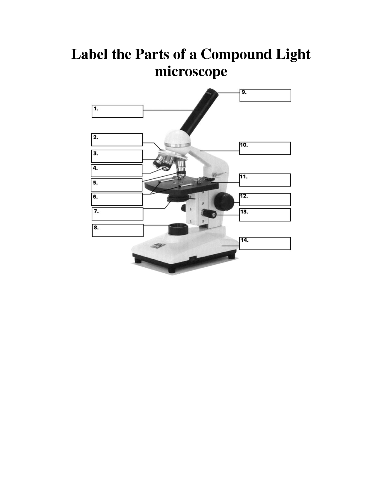
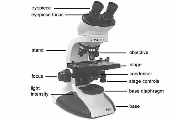
rsscience.com › stereo-microscopeParts of Stereo Microscope (Dissecting microscope) - Rs' Science Optical parts of a stereo microscope work together to magnify and produce a 3-D image of the specimens. These parts include: Eyepieces. The eyepiece (or ocular lens) is the lens part at the top of a microscope that the viewer looks through. Typically, standard eyepieces for a dissecting microscope have a magnifying power of 10x.

Human Eye Ball Anatomy & Physiology Diagram - eMedicineHealth The cornea is located just in front of the iris, which is the colored part of the eye. The main purpose of the cornea is to help focus light as it enters the eye. If one wears contact lenses, the contact lens rests on the cornea. Iris and Pupil. The iris, which is the colored part of the eye, controls the amount of light that enters the eye.

Activity Sheet 1: How the Eyes Work | Human eye diagram, Teaching biology, Human body activities
› engine-vu › yamaha-marine-genuineComplete Yamaha Marine Outboard OEM Parts Catalog | PartsVu Yamaha outboard parts are the heart and soul of PartsVu. Offering more than 40,000 Genuine Yamaha outboard parts and products and at low prices with same business day shipping on in-stock items. We value convenience and knowledge, providing resources like our Yamaha Outboard Maintenance Parts Charts , helping you to find the right parts every time.

Eye Diagram Without Labels | via Anatomy Pictures Gallery if… | Flickr
Eye Anatomy: Parts of the Eye and How We See Behind the anterior chamber is the eye's iris (the colored part of the eye) and the dark hole in the middle called the pupil. Muscles in the iris dilate (widen) or constrict (narrow) the pupil to control the amount of light reaching the back of the eye. Directly behind the pupil sits the lens. The lens focuses light toward the back of the eye.
Solved: Label the parts of the eye: | Chegg.com
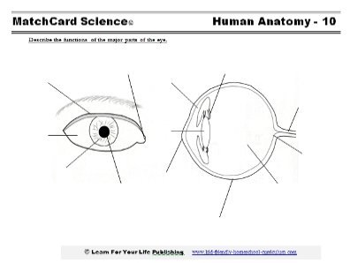
Label the Eye - The Biology Corner Label the Eye. Shannan Muskopf December 30, 2019. This worksheet shows an image of the eye with structures numbered. Students practice labeling the eye or teachers can print this to use as an assessment. There are two versions on the google doc and pdf file, one where the word bank is included and another with no word bank for differentiation.

Label the Eye
The Eyes (Human Anatomy): Diagram, Optic Nerve, Iris, Cornea ... - WebMD The front part (what you see in the mirror) includes: Iris: the colored part. Cornea: a clear dome over the iris. Pupil: the black circular opening in the iris that lets light in. Sclera: the ...
The Parts of the Eye - TeacherVision
Parts of the Eye & Their Function | Robertson Optical and Optometry The different parts of the eye allow the body to take in light and perceive objects around us in the proper color, detail and depth. This allows people to make more informed decisions about their environment. If a portion of the eye becomes damaged, you may not be able to see effectively, or lose your vision all together.

Eye Diagram With Labels and detailed description - BYJUS Iris is the coloured part of the eye and controls the amount of light entering the eye by regulating the size of the pupil. The lens is located just behind the iris. Its function is to focus the light on the retina. The optic nerve transmits electrical signals from the retina to the brain. Pupil is the opening at the centre of the iris.

Parts of the Eye - Primary copy.pdf
Eye Anatomy Detail Picture Image on MedicineNet.com Picture of Eye Anatomy Detail The eye is our organ of sight. The eye has a number of components which include but are not limited to the cornea, iris, pupil, lens, retina, macula, optic nerve, choroid and vitreous. Cornea: clear front window of the eye that transmits and focuses light into the eye.
Human Eye Diagram
Cornea of the Eye - Definition and Detailed Illustration Cornea Definition. The cornea is the clear front surface of the eye. It lies directly in front of the iris and pupil, and it allows light to enter the eye. Viewed from the front of the eye, the cornea appears slightly wider than it is tall. This is because the sclera (the "white" of the eye) slightly overlaps the top and bottom of the anterior ...
8 Best Images of Lens Diagram Worksheet - Microscope with Labeled Parts, Label Eye Parts ...
Packaging | Custom Boxes Wholesale | Create Your Own ... Finding custom boxes online is easy, but ordering the right custom boxes without experience is incredibly hard. At Emenac Packaging, real packaging experts poring over the details to ensure perfection. They are available 24/7 to assist you choose the right stock, size, colors, and quantity to help process your order faster and more reliable. Here.
31 Eye Label Quiz - Labels 2021
idlabelinc.com › common-types-warehouse-labelsCommon Types of Warehouse Labels - ID Label Inc. Warehouse Tote and Bin Labels. Warehouses commonly store individual products and parts in plastic bins or containers. Like warehouse racks, these bins should be properly identified with barcode labels to help workers easily locate items, fulfill orders and manage product inventory. Similarly, reusable warehouse totes require proper identification.
8 Best Images of Lens Diagram Worksheet - Microscope with Labeled Parts, Label Eye Parts ...
Anatomy of the eye: Quizzes and diagrams - Kenhub Found within two cavities in the skull known as the orbits, the eyes are surrounded by several supporting structures including muscles, vessels, and nerves. There are 7 bones of the orbit, two groups of muscles (intrinsic ocular and extraocular), three layers to the eyeball … and that's just the beginning. There's a lot to learn, but stay calm!

Learn the Nine Essential Parts of Eyeglasses 1. Rims The rims lend form and character to your eyeglasses—they also provide function by holding the lenses in place. 2. End pieces The end pieces are the small parts on the frame that extend outward and connect the lenses to the temples. 3. Bridge The bridge is the center of the frame that rests on your nose and joins the two rims together. 4.

30 Label The Parts Of Eye - Label Design Ideas 2020
Label Parts of the Human Ear - University of Dayton Label Parts of the Human Ear. Select One Auditory Canal Cochlea Cochlear Nerve Eustachian Tube Incus Malleus Oval Window Pinna Round Window Semicircular Canals Stapes Tympanic Membrane Vestibular Nerve. Select One Auditory Canal Cochlea Cochlear Nerve Eustachian Tube Incus Malleus Oval Window Pinna Round Window Semicircular Canals Stapes ...

31 Parts Of The Eye With Label - Label Design Ideas 2020
Parts of the Eye - RIT Parts of the Eye Here I will briefly describe various parts of the eye: Sclera The sclera is the white of the eye. "Don't shoot until you see their scleras." Exterior is smooth and white Interior is brown and grooved Extremely durable Flexibility adds strength Continuous with sheath of optic nerve Tendons attached to it The Cornea
Eyes - PowerKnowledge Life Science
reference.yourdictionary.com › resources › parts-ofParts of the Body for Kids: Names & Basic Functions Diagram of Body Parts. External, which means “outside,” describes the body parts that you can see. Take a look at a helpful diagram that labels major external body parts. Download the printable PDF to see it in more detail and print if needed. View & Download PDF
Post a Comment for "44 parts of the eye without labels"