40 diagram of human heart with labels
File:Diagram of the human heart (cropped).svg - Wikimedia Aug 08, 2022 · English: Diagram of the human heart 1. Superior vena cava 2. 4. Mitral valve 5. Aortic valve 6. Left ventricle 7. Right ventricle 8. Left atrium 9. Right atrium 10. Aorta 11. Pulmonary v 14,796 Human heart diagram Images, Stock Photos & Vectors - Shutterstock 14,796 human heart diagram stock photos, vectors, and illustrations are available royalty-free. See human heart diagram stock video clips Image type Orientation Color People Artists Sort by Popular Anatomy Healthcare and Medical Abstract Designs and Shapes heart medicine organ circulatory system hemodynamics cardiology vein Next of 148
How to Draw a Human Heart: 11 Steps (with Pictures) - wikiHow Make an oval that touches each point of the triangle. Then, draw the main part of the heart within them. 2. Make a rounded bump at the top of the heart for the right atrium. Draw a half-circle or bump that extends from the top left corner of the heart.
Diagram of human heart with labels
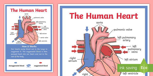
A Labeled Diagram of the Human Heart You Really Need to See The human heart, comprises four chambers: right atrium, left atrium, right ventricle and left ventricle. The two upper chambers are called the left and the right atria, and the two lower chambers are known as the left and the right ventricles. The two atria and ventricles are separated from each other by a muscle wall called 'septum'. Human Heart (Anatomy): Diagram, Function, Chambers, Location ... Cardiomyopathy: A disease of heart muscle in which the heart is abnormally enlarged, thickened, and/or stiffened. As a result, the heart's ability to pump blood is weakened. As a result, the heart ... Human Heart Diagram Class 10 | Get Easy Tricks to Draw Human Heart Human Heart Diagram with Label. In your exams, your diagram will be marked only if you have labeled it. That is how important labels are. If you practice the image multiple times with the labels, you will automatically remember all the marking labels. And to add to it, you must make sure all your marking labels are aligned.
Diagram of human heart with labels. Human Heart Diagram Labeled | Science Trends Let's examine the anatomy of the heart along with some diagrams that show how the heart operates. Anatomy Of The Heart The human heart usually weighs somewhere between 10 to 12 ounces in men and between 8 to 10 ounces in women, and in terms of size is roughly the size of the fist. Free Printable Heart Diagram for Kids - Labeled and Unlabeled Oct 17, 2018 - Learn all the parts of the human heart by memorizing this free printable human heart diagram. Includes labeled and unlabeled versions. Pinterest. Today. Explore. When autocomplete results are available use up and down arrows to review and enter to select. Touch device users, explore by touch or with swipe gestures. Heart Anatomy: Labeled Diagram, Structures, Blood Flow ... - EZmed There are 4 chambers, labeled 1-4 on the diagram below. To help simplify things, we can convert the heart into a square. We will then divide that square into 4 different boxes which will represent the 4 chambers of the heart. The boxes are numbered to correlate with the labeled chambers on the cartoon diagram. View fullsize Label the heart — Science Learning Hub In this interactive, you can label parts of the human heart. Drag and drop the text labels onto the boxes next to the diagram. Selecting or hovering over a box will highlight each area in the diagram. pulmonary vein semilunar valve right ventricle right atrium vena cava left atrium pulmonary artery aorta left ventricle Download Exercise Tweet
Human Heart Diagram - Human Body Pictures - Science for Kids Find free pictures, photos, diagrams, images and information related to the human body right here at Science Kids. Photo name: Human Heart Diagram Picture category: Human Body Image size: 70 KB Dimensions: 600 x 600 Photo description: This is an excellent human heart diagram which uses different colors to show different parts and also labels a number of important heart component such as the ... The Human Heart Labeling Worksheet (Teacher-Made) - Twinkl The human heart is a muscle made up of four chambers, these are: Two upper chambers - the left atrium and right atrium Two lower chambers - the left and right ventricles. It's also made up of four valves - these are known as the tricuspid, pulmonary, mitral and aortic valves. Heart Diagram | Free Heart Diagram Templates - Edrawsoft Do you know that human heart system can be even more powerful than an electronic equipment? Wanna figure out why? ... Just refer to this originally designed Edraw heart diagram science template for more details. Lab Apparatus List. 64704. 211. Plant Cell Diagram. 19550. 173. Heart Diagram. 18805. 156. Food Web Diagram. 11966. 154. Leaf Cross ... Human Heart - Diagram and Anatomy of the Heart - Innerbody Chambers of the Heart The heart contains 4 chambers: the right atrium, left atrium, right ventricle, and left ventricle. The atria are smaller than the ventricles and have thinner, less muscular walls than the ventricles. The atria act as receiving chambers for blood, so they are connected to the veins that carry blood to the heart.
Human Heart Diagram - Side View and Top View - Heart Valve Surgery As shown below, this human heart diagram clearly illustrates the valves of the heart. The valves illustrated below are the pulmonary, tricuspid, aortic and mitral valve. So you know, I had the aortic and pulmonary valves of my heart replaced via the Ross Procedure. Understanding Human Heart with Heart Diagram | EdrawMax Online - Edrawsoft Step 1: Open EdrawMax, and select Science and Education, then click on human organs. Source: EdrawMax. Step 2: Use the wide range of symbols from the libraries available to create your heart diagram. Source: EdrawMax. Step 3: Add in your text and label the heart diagram to suit the requirements. Male Human Anatomy Diagram Pictures, Images and Stock Photos Pacemaker Diagram Cross section of a human heart with pacemaker fitted, showing the major arteries and veins. This is an EPS 10 vector illustration and includes a high resolution JPEG. male human anatomy diagram stock illustrations Blood Flow Through The Heart: A Simple 12 Step Diagram - EZmed Step 1 and 6 involve a blood vessel, which makes sense as this is how blood enters and exits that side of the heart. Steps 2-5 involve a chamber, valve, chamber, and valve. So if you remember this general pattern, it will help you recall the order in which blood flows through each side of the heart.
File:Diagram of the human heart (no labels).svg File:Diagram of the human heart (no labels).svg. From Wikimedia Commons, the free media repository. File. File history. File usage on Commons. Metadata. Size of this PNG preview of this SVG file: 498 × 599 pixels. Other resolutions: 199 × 240 pixels | 399 × 480 pixels | 639 × 768 pixels | 851 × 1,024 pixels | 1,703 × 2,048 pixels | 533 × ...
Diagram of Human Heart and Blood Circulation in It Exterior of the Human Heart A heart diagram labeled will provide plenty of information about the structure of your heart, including the wall of your heart. The wall of the heart has three different layers, such as the Myocardium, the Epicardium, and the Endocardium. Here's more about these three layers. Epicardium
File:Diagram of the human heart (cropped).svg - Wikipedia Diagram of the human heart, created by Wapcaplet in Sodipodi. Cropped by Yaddah to remove white space ... Add Inferior vena cava and pericardium labels: 18:08, 14 August 2018: 656 × 631 (209 KB) Jmarchn: Add pericardium. Add papillary muscles and chordae tendinae. Add cardiac skeleton. Inferior vena cava more wide.
Pin on Anatomy - Pinterest yd941uzen: arteries of heart diagram. Find this Pin and more on Anatomy by Tiffany Riddick. Subclavian Artery. Carotid Artery. Heart Pictures. Heart Images. Heart Blood Flow. Human Heart Diagram. Heart Structure.
Human Heart Diagram Pictures, Images and Stock Photos A medical diagram showing the heart, arteries and veins of the human body. Cross Section of Heart with Labels on White Background Computer generated image of a sagittal cross section view of a human heart, showing chambers, major arteries and veins with anatomy labels. 3d rendering of the human heart anatomy Human heart angioplasty
Diagram of the human heart royalty-free images - Shutterstock 14,791 diagram of the human heart stock photos, vectors, and illustrations are available royalty-free. See diagram of the human heart stock video clips Image type Orientation Color People Artists Sort by Popular Anatomy Healthcare and Medical Icons and Graphics Diseases, Viruses, and Disorders heart medicine organ hemodynamics circulatory system

QuickStudy | Nervous System Laminated Study Guide | Nervous system, Heart attack symptoms, Anatomy
Human Heart - Anatomy, Functions and Facts about Heart - BYJUS The human heart is one of the most important organs responsible for sustaining life. It is a muscular organ with four chambers. The size of the heart is the size of about a clenched fist. The human heart functions throughout a person’s lifespan and is one of the most robust and hardest working muscles in the human body.

Detailed Labeled Anatomy Human Body | jpg: labeled heart flow | Nursing | Pinterest | Heart ...
Heart Diagram – 15+ Free Printable Word, Excel, EPS, PSD ... Teachers and students use the heart diagram, in biological science, to study the structure and functions of a human being’s heart. Friends and colleagues on the other hand may find this diagram template useful when it comes to sending special, personalized gifts to their family members and significant others. Download the template today, and ...
13+ Heart Diagram Templates - Sample, Example, Format Download Color Heart Diagram Sample Format Free Download. cdhb.health.nz This colored heart diagram is a graphic representation of the organ which can be used for presentations and videos about the subject of human heart. The picture is in a coloured format and is available for a free download. Free Download.
A Diagram of the Heart and Its Functioning Explained in Detail The heart blood flow diagram (flowchart) given below will help you to understand the pathway of blood through the heart.Initial five points denotes impure or deoxygenated blood and the last five points denotes pure or oxygenated blood. 1.Different Parts of the Body ↓ 2.Major Veins ↓ 3.Right Atrium ↓ 4.Right Ventricle ↓ 5.Pulmonary Artery ↓ 6.Lungs
Human Heart with Labels on White Background stock photo ... Description Computer generated image of human heart, major arteries and veins with labeled anatomy, isolated on white background 1 credit Essentials collection for this image $4 with a 1-month subscription (10 Essentials images for $40) Continue with purchase View plans and pricing Includes our standard license. Add an extended license.
How to Draw the Internal Structure of the Heart (with Pictures) - wikiHow To draw the internal structure of a human heart, follow the steps below. Part 1 Finding a Diagram 1 To find a good diagram, go to Google Images, and type in "The Internal Structure of the Human Heart". Find an image that displays the entire heart, and click on it to enlarge it. 2 Find a piece of paper and something to draw with.
Heart Diagram with Labels and Detailed Explanation - BYJUS The human heart is the most crucial organ of the human body. It pumps blood from the heart to different parts of the body and back to the heart. The most common heart attack symptoms or warning signs are chest pain, breathlessness, nausea, sweating etc. The diagram of heart is beneficial for Class 10 and 12 and is frequently asked in the ...
Human Heart Diagram Class 10 | Get Easy Tricks to Draw Human Heart Human Heart Diagram with Label. In your exams, your diagram will be marked only if you have labeled it. That is how important labels are. If you practice the image multiple times with the labels, you will automatically remember all the marking labels. And to add to it, you must make sure all your marking labels are aligned.

A Labeled Diagram of the Human Heart You Really Need to See | Human heart and Nursing students
Human Heart (Anatomy): Diagram, Function, Chambers, Location ... Cardiomyopathy: A disease of heart muscle in which the heart is abnormally enlarged, thickened, and/or stiffened. As a result, the heart's ability to pump blood is weakened. As a result, the heart ...
the major blood vessels of heart using interactive animations diagrams labels the Major Blood ...
A Labeled Diagram of the Human Heart You Really Need to See The human heart, comprises four chambers: right atrium, left atrium, right ventricle and left ventricle. The two upper chambers are called the left and the right atria, and the two lower chambers are known as the left and the right ventricles. The two atria and ventricles are separated from each other by a muscle wall called 'septum'.


Post a Comment for "40 diagram of human heart with labels"